The Critical Role of Image Resolution in Revealing Microscopic Particl…
페이지 정보

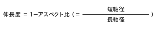
본문
Pixel density plays a critical role in capturing sub-micron features, particularly in industrial imaging applications where clarity is paramount. Image sharpness refers to the number of distinct pixels in an image, typically expressed as horizontal by vertical dimensions, and directly influences the level of detail that can be resolved. When observing particles that are sub-micrometer or near the limits of human vision, enhanced spatial sampling becomes mandatory to distinguish distinct surface topographies, microscopic roughness, and subtle variations in shape or density.
In fields such as materials science, air quality analysis, and drug formulation, particles can range from a few micrometers down to atomic-scale sizes. A blurry capture may depict these particles as unresolved artifacts, making it impossible to accurately count, segment, or evaluate them. Precision optical sensors, by contrast, provide adequate sampling rate to isolate single entities and even submicron anomalies such as pores, cracks, or surface layers. This enhanced definition enables researchers to detect anomalies, quantify size spectra with reduced error margins, and observe dynamic behavior under varying conditions.
The interplay of magnification and pixel density is also crucial. Increasing enlargement without a corresponding increase in resolution leads to enlarged but blurry images, a phenomenon commonly referred to as empty magnification. Accurate feature resolution requires both proper focal extension and a detector with sufficient sampling the sub-pixel details being observed. This is why advanced optical systems, SEM, and scientific CMOS cameras are designed for high pixel counts, high signal-to-noise ratio detectors, and 粒子径測定 precision optics optimized for fine detail.
Moreover, image sharpness affects the reliability of algorithmic processing. Many automated inspection systems rely on algorithms to identify and quantify particles. These computational models depend on clear boundaries and stable intensity differentials between particles and their background. Low-resolution images introduce uncertainty, leading to false detections, missed detections, or quantification errors. Sharp, detailed imagery ensures that machine learning models can operate with higher accuracy and enhanced reproducibility.
It is also important to consider the physical boundaries imposed by the physical properties of light. In conventional microscopy, Abbe limit restricts the resolution threshold to approximately half the wavelength of the illumination source. To extend beyond this limit, techniques such as STORM have been refined, pushing the limits of observable detail and still maintaining biological relevance. These next-generation imaging protocols still rely on ultra-sensitive cameras to digitize the refined data produced by the optical setup.
In real-world applications, choosing the optimal pixel density involves optimizing image quality with file size, processing speed, and system investment. While higher resolution yields richer information, it also generates larger file sizes and demands more computational power. For quality control checks, where only general particle sizes are needed, baseline imaging may suffice. But for research involving molecular interactions, pollutant detection, or material failure analysis, only high-resolution imaging can provide the actionable data.
Ultimately, the ability to capture nanoscale structures hinges on the suitability and fidelity of the pixel sampling. Without proper sampling, even the cutting-edge software cannot make up for unresolved features. Deploying appropriate microscopy systems ensures that key micro-features are preserved, enabling validated results, strategic insights, and enhanced knowledge.
- 이전글Advanced Dynamic Imaging Techniques for Resin Granule Analysis 25.12.31
- 다음글บาคาร่า 25.12.31
댓글목록
등록된 댓글이 없습니다.